
CAP management begins with severity stratification — not empiric antibiotic selection. The level of care decision, antibiotic regimen, and monitoring intensity all follow from an accurate severity assessment. Atypical pathogens require specific coverage, and radiographic lag is the rule, not the exception.
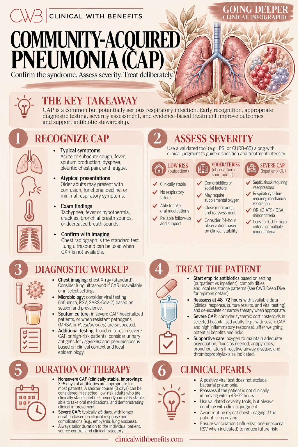
1 · Diagnosis — Clinical, Radiographic, and Laboratory
Community-acquired pneumonia is diagnosed by the combination of a new pulmonary infiltrate on chest imaging plus clinical signs and symptoms of lower respiratory tract infection — fever, cough, sputum production, pleuritic chest pain, dyspnea, or abnormal lung exam findings. No single finding is sufficient; the diagnosis requires integrating the clinical picture with imaging.
Chest X-ray is the standard initial imaging study. A new infiltrate — consolidation, interstitial pattern, or mixed pattern — in the appropriate clinical context confirms the diagnosis. CT chest is more sensitive than plain X-ray and is appropriate when the diagnosis is uncertain, when the patient is not improving as expected, or when a complication (empyema, lung abscess) is suspected. A normal chest X-ray in a patient with clinical signs of pneumonia does not rule out early CAP — repeat imaging at 24–48 hours may be appropriate in high-suspicion cases.
Laboratory findings in CAP are supportive but not diagnostic. Leukocytosis with a left shift is common but nonspecific. Procalcitonin is elevated in bacterial pneumonia and can help distinguish bacterial from viral lower respiratory tract infection — but it should not be used to withhold antibiotics when the clinical and radiographic picture is consistent with CAP. Blood cultures are recommended before antibiotics in hospitalized patients, particularly those with severe CAP or those who may have bacteremic pneumonia. Sputum Gram stain and culture are low yield in most outpatient and non-ICU inpatient settings but are appropriate in severe CAP.
2 · Severity Assessment — CURB-65 and PSI/PORT
Severity assessment is the first and most important step in CAP management — it determines the level of care, antibiotic regimen, and monitoring intensity. Two validated scoring systems are widely used: CURB-65 and the Pneumonia Severity Index (PSI/PORT).
CURB-65 assigns one point each for: Confusion (new disorientation), Urea above 7 mmol/L (BUN above 19 mg/dL), Respiratory rate 30 or above, Blood pressure below 90 mmHg systolic or 60 mmHg diastolic, and age 65 or older. Score 0–1: low risk, outpatient treatment appropriate in most patients. Score 2: intermediate risk, consider hospitalization. Score 3 or higher: high risk, hospitalization required; score 4–5 warrants consideration of ICU-level care.
PSI/PORT stratifies patients into five risk classes based on age, comorbidities, physical exam findings, and laboratory values. Risk classes I–II support outpatient treatment; class III may be treated outpatient or with brief hospitalization; classes IV–V require hospitalization. PSI/PORT is more complex to calculate than CURB-65 but is more discriminating at the low end of severity — it identifies more patients who can safely be treated outpatient.
Both scoring systems are tools to inform clinical judgment, not replace it. A patient with a low CURB-65 score who has poor social support, inability to take oral medications, or significant comorbidities may still require hospitalization. Clinical gestalt and the ability to follow up reliably are part of the disposition decision.
3 · Antibiotic Selection — Empiric Regimens by Severity
Empiric antibiotic selection in CAP is guided by severity, comorbidities, and risk factors for resistant organisms. The IDSA/ATS 2019 guidelines provide the current evidence-based framework.
Outpatient treatment, no comorbidities: amoxicillin 1 g three times daily is the preferred agent for otherwise healthy patients without comorbidities. Doxycycline 100 mg twice daily is an alternative. Azithromycin monotherapy is no longer recommended as first-line in areas with high macrolide resistance rates (above 25%).
Outpatient treatment, with comorbidities (chronic heart, lung, liver, or renal disease; diabetes; immunosuppression; use of antimicrobials in the past 3 months): combination therapy with amoxicillin-clavulanate or a cephalosporin plus a macrolide, or respiratory fluoroquinolone monotherapy (levofloxacin 750 mg daily or moxifloxacin 400 mg daily).
Inpatient, non-ICU: combination therapy with a beta-lactam (ampicillin-sulbactam, ceftriaxone, or cefotaxime) plus a macrolide (azithromycin or clarithromycin), or respiratory fluoroquinolone monotherapy. The beta-lactam + macrolide combination is preferred over fluoroquinolone monotherapy in most patients because it provides better coverage for Streptococcus pneumoniae and has a more favorable resistance profile.
Inpatient, ICU (severe CAP): beta-lactam plus azithromycin, or beta-lactam plus a respiratory fluoroquinolone. For patients with Pseudomonas risk factors (structural lung disease, recent hospitalization, prior Pseudomonas isolation, immunosuppression), use an anti-pseudomonal beta-lactam (piperacillin-tazobactam, cefepime, or meropenem) plus a fluoroquinolone or aminoglycoside plus azithromycin.
4 · Atypical Pathogens — Mycoplasma, Chlamydophila, and Legionella
Atypical pathogens — Mycoplasma pneumoniae, Chlamydophila pneumoniae, and Legionella pneumophila — cause a significant proportion of CAP and do not respond to beta-lactam antibiotics. This is why beta-lactam monotherapy is insufficient for most hospitalized CAP patients and why atypical coverage (macrolide or fluoroquinolone) is required.
Mycoplasma pneumoniae is the most common atypical pathogen in younger patients and outpatient CAP. It typically causes a milder illness — often called "walking pneumonia" — with gradual onset, dry cough, and low-grade fever. Chest X-ray may show a patchy interstitial pattern. Macrolides and doxycycline are effective; fluoroquinolones are alternatives.
Legionella pneumophila causes a more severe illness — Legionnaires' disease — and should be specifically considered in patients with severe CAP, hyponatremia (a classic associated finding), diarrhea, recent travel (particularly hotel stays or cruise ships), exposure to water systems (cooling towers, hot tubs), or immunocompromised states. Urine Legionella antigen is the most practical diagnostic test and detects serogroup 1, which causes the majority of severe disease. Respiratory fluoroquinolones and azithromycin are the treatments of choice — beta-lactams are ineffective.
Procalcitonin can help distinguish bacterial from viral lower respiratory tract infection — a low procalcitonin (below 0.1 ng/mL) in a patient with mild symptoms and no radiographic infiltrate suggests a viral etiology and supports withholding antibiotics. However, atypical pathogens can cause CAP with relatively low procalcitonin levels. In a patient with a clear infiltrate and clinical signs of pneumonia, antibiotics should not be withheld based on procalcitonin alone.
5 · Expected Recovery and Radiographic Lag
The expected recovery timeline for CAP is predictable. Fever and leukocytosis typically improve within 2–4 days of starting appropriate antibiotics. Clinical improvement — reduced dyspnea, improved oxygenation, ability to tolerate oral medications — usually occurs within 3–5 days. Changing antibiotics at 48 hours because the patient still has a fever is premature and inappropriate.
Chest X-ray infiltrates lag significantly behind clinical improvement. Infiltrates can persist 4–6 weeks after clinical resolution, particularly in older patients and those with underlying lung disease. A chest X-ray that shows persistent infiltrates at 48–72 hours is not evidence of treatment failure — it is the expected radiographic course. Follow-up chest X-ray at 6–8 weeks after discharge is recommended to confirm clearance and exclude an underlying mass (particularly in smokers and older patients).
True treatment failure is defined by clinical deterioration — worsening oxygenation, hemodynamic instability, failure to defervesce after 72 hours, or development of new complications. When treatment failure occurs, the differential includes: resistant organisms (MRSA, Pseudomonas in patients without appropriate coverage), atypical coverage gaps (Legionella not covered by beta-lactam monotherapy), empyema, lung abscess, non-infectious mimics (pulmonary edema, malignancy, organizing pneumonia, pulmonary hemorrhage), and immunocompromised states.
6 · Complications — Empyema, Lung Abscess, and Respiratory Failure
Empyema — infected pleural fluid — is the most important complication to recognize because it requires drainage in addition to antibiotics. Antibiotics alone will not resolve an empyema. Clinical clues include persistent fever despite appropriate antibiotics, pleuritic chest pain, and a new or enlarging pleural effusion. CT chest with contrast is the most sensitive imaging study. Thoracentesis with pleural fluid analysis (pH, glucose, LDH, Gram stain, culture) confirms the diagnosis. Drainage — thoracentesis, chest tube, or surgical decortication depending on the stage — is required.
Lung abscess results from necrotizing pneumonia with cavitation. It is more common with aspiration pneumonia, anaerobic organisms, and immunocompromised states. Chest X-ray or CT shows a cavitary lesion, often with an air-fluid level. Treatment requires prolonged antibiotics (4–6 weeks or until the cavity resolves) with anaerobic coverage (amoxicillin-clavulanate, clindamycin, or a carbapenem). Most lung abscesses resolve with antibiotics alone; drainage is reserved for abscesses that fail to respond.
Respiratory failure in severe CAP requires escalating respiratory support. High-flow nasal cannula (HFNC) is appropriate for hypoxemic respiratory failure not responding to conventional oxygen — it reduces the need for intubation in non-hypercapnic patients. NIV (BiPAP) is less well-established in CAP than in COPD exacerbation, but may be appropriate in selected patients. Early intubation should not be delayed in patients with severe CAP and worsening respiratory failure — delayed intubation is associated with worse outcomes.
7 · Discharge Criteria, Duration, and Follow-Up
Discharge criteria for hospitalized CAP: clinical stability — temperature below 37.8°C, heart rate below 100, respiratory rate below 24, systolic blood pressure above 90 mmHg, oxygen saturation above 90% on room air or baseline supplemental oxygen, ability to maintain oral intake, and normal mental status. Patients who meet clinical stability criteria can be safely discharged on oral antibiotics.
Antibiotic duration: 5 days is appropriate for most patients with CAP who achieve clinical stability. The IDSA/ATS guidelines recommend a minimum of 5 days; longer courses are appropriate for patients with bacteremia, Legionella, or complications. Procalcitonin-guided de-escalation can safely reduce antibiotic duration in hospitalized CAP patients.
Follow-up: outpatient follow-up within 1–2 weeks of discharge is recommended. Repeat chest X-ray at 6–8 weeks confirms radiographic clearance and excludes an underlying mass. Pneumococcal vaccination status should be reviewed at follow-up — patients with CAP who are not up to date on pneumococcal vaccines should receive them. Influenza vaccination should also be reviewed.
Apply It · Patient Scenario
A 68-year-old man with COPD and type 2 diabetes presents with 4 days of productive cough, fever to 38.9°C, and dyspnea. He is confused on exam. Respiratory rate is 28. Blood pressure is 96/58 mmHg. SpO2 is 88% on room air. Chest X-ray shows a right lower lobe consolidation. BUN is 24 mg/dL. He is admitted to the floor and started on ceftriaxone monotherapy.
What is the most important problem with the current management?
A. The patient should have been treated outpatient — his CURB-65 score is only 2
B. Ceftriaxone monotherapy does not cover atypical pathogens — a macrolide or respiratory fluoroquinolone should be added, and the severity score suggests ICU-level care should be considered
C. The chest X-ray should be repeated in 48 hours to confirm the infiltrate is resolving before starting antibiotics
D. Procalcitonin should be checked before starting antibiotics to confirm bacterial etiology
ANSWER
B. Ceftriaxone monotherapy is insufficient — atypical coverage is required, and the CURB-65 score indicates high severity.
RATIONALE
This patient's CURB-65 score is 4: Confusion (1) + BUN above 19 mg/dL (1) + Respiratory rate 28 — below the ≥30 threshold, so 0 points — + Blood pressure below 90 systolic (1) + Age 68 (1). A CURB-65 of 3 or higher indicates high severity and requires hospitalization; a score of 4 places this patient at the high end of that range. CURB-65 alone does not determine ICU disposition — that decision requires applying the IDSA/ATS severe CAP criteria (one major criterion: septic shock requiring vasopressors or respiratory failure requiring mechanical ventilation; or three or more minor criteria including RR ≥30, PaO2/FiO2 ≤250, multilobar infiltrates, confusion, BUN ≥20 mg/dL, leukopenia, thrombocytopenia, hypothermia, or hypotension requiring aggressive resuscitation). This patient's hypotension, confusion, and hypoxia warrant urgent reassessment against those criteria before disposition is finalized.
Ceftriaxone monotherapy does not cover Mycoplasma, Chlamydophila, or Legionella. For hospitalized non-ICU CAP, IDSA/ATS guidelines recommend beta-lactam plus macrolide or respiratory fluoroquinolone monotherapy. For severe CAP (ICU-level), beta-lactam plus azithromycin or beta-lactam plus respiratory fluoroquinolone is required. Legionella should be specifically considered given the severity — urine Legionella antigen should be sent.
Waiting for a repeat chest X-ray before starting antibiotics is incorrect — antibiotics should be started promptly in hospitalized CAP. Procalcitonin does not determine whether to start antibiotics in a patient with a clear infiltrate and clinical signs of pneumonia.
Clinical Pearl: CURB-65 ≥3 requires hospitalization. ICU disposition is determined by IDSA/ATS severe CAP criteria — not CURB-65 alone. Beta-lactam monotherapy is insufficient for hospitalized CAP — always add atypical coverage.
NOW CHANGE ONE DETAIL
Same patient. He is admitted to the ICU, started on ceftriaxone plus azithromycin, and improves over 48 hours. On day 4, he develops worsening fever, increasing oxygen requirements, and a new right-sided pleural effusion on chest X-ray.
UPDATED REASONING
A new or enlarging pleural effusion in a patient with CAP who was initially improving should raise immediate concern for empyema. Antibiotics alone will not resolve an empyema — drainage is required. The next step is CT chest with contrast to characterize the effusion, followed by thoracentesis with pleural fluid analysis (pH, glucose, LDH, Gram stain, culture). A pH below 7.2, glucose below 60 mg/dL, or positive Gram stain/culture confirms empyema and mandates drainage.
The lesson: clinical deterioration after initial improvement in CAP should prompt a systematic search for complications — empyema, lung abscess, resistant organisms, and non-infectious mimics — rather than empiric antibiotic escalation alone.
Understand It · The Nuance
The most common errors in CAP management are undertreating severity, omitting atypical coverage, changing antibiotics prematurely for radiographic lag, and missing empyema when treatment fails.
Severity assessment drives every decision
CAP management begins with severity stratification — not empiric antibiotic selection. PSI/PORT and CURB-65 are validated tools that guide the outpatient vs. inpatient vs. ICU decision. A CURB-65 score of 0–1 supports outpatient treatment in most patients; a score of 3 or higher warrants hospitalization and consideration of ICU-level care.
Atypical coverage is not optional in most hospitalized patients
Mycoplasma pneumoniae, Chlamydophila pneumoniae, and Legionella pneumophila cause a significant proportion of CAP and do not respond to beta-lactams alone. IDSA/ATS guidelines recommend combination therapy (beta-lactam + macrolide) or respiratory fluoroquinolone monotherapy for hospitalized non-ICU patients. Legionella should be specifically considered in severe CAP, immunocompromised patients, and patients with hyponatremia, diarrhea, or recent travel.
Procalcitonin guides antibiotic duration, not initiation
In CAP, procalcitonin is most useful for guiding antibiotic de-escalation and duration — not for deciding whether to start antibiotics. A patient with clinical, radiographic, and laboratory evidence of CAP should receive antibiotics regardless of procalcitonin level. Serial procalcitonin trending supports earlier discontinuation in patients who are improving.
Radiographic findings lag behind clinical improvement
Chest X-ray infiltrates can persist 4–6 weeks after clinical resolution of CAP. Persistent radiographic findings at 48–72 hours are not treatment failure. True treatment failure is defined by clinical deterioration — worsening oxygenation, hemodynamic instability, or failure to defervesce — not by persistent infiltrates on imaging.
When CAP is not responding, broaden the differential
Failure to improve with standard CAP treatment should prompt consideration of resistant organisms, atypical coverage gaps, empyema, lung abscess, non-infectious mimics (pulmonary edema, malignancy, organizing pneumonia), and immunocompromised states. Empyema requires drainage — antibiotics alone will not resolve it.
Oxygenation targets matter
Target SpO2 92–96% in most CAP patients. In patients with known COPD or chronic hypercapnia, target 88–92% to avoid suppressing hypoxic ventilatory drive. High-flow nasal cannula is appropriate for hypoxemic respiratory failure not responding to conventional oxygen. Early escalation to NIV or intubation should not be delayed in patients with severe CAP and respiratory failure.
Clinical Pearl: Chest X-ray infiltrates can persist 4–6 weeks after clinical resolution. Do not change antibiotics at 48 hours because the X-ray looks the same — true treatment failure is clinical deterioration, not radiographic persistence.
Clinical Pearls
Clinical Pearls
- Severity Score FirstCURB-65 and PSI/PORT guide the outpatient vs. inpatient vs. ICU decision. A CURB-65 of 0–1 supports outpatient treatment in most patients; 3 or higher warrants hospitalization.
- Atypical Coverage Is RequiredBeta-lactam monotherapy is insufficient for most hospitalized CAP. Add a macrolide or use a respiratory fluoroquinolone to cover Mycoplasma, Chlamydophila, and Legionella.
- Think Legionella in Severe CAPLegionella should be specifically considered in severe CAP, hyponatremia, diarrhea, recent travel, or immunocompromised patients. Urine Legionella antigen detects serogroup 1 (the most common cause of severe disease).
- Radiographic Lag Is NormalChest X-ray infiltrates can persist 4–6 weeks after clinical resolution. Persistent infiltrates at 48–72 hours are not treatment failure. True failure is clinical deterioration.
- Procalcitonin Guides DurationProcalcitonin is most useful for guiding antibiotic de-escalation and duration in CAP — not for deciding whether to start antibiotics. Start antibiotics based on clinical and radiographic evidence.
- Empyema Requires DrainageWhen CAP is not responding, consider empyema. Antibiotics alone will not resolve an empyema — drainage is required. CT chest or ultrasound-guided thoracentesis is the next step.
Bottom Line
CAP management begins with severity stratification — CURB-65 or PSI/PORT determines level of care. Atypical coverage is required for most hospitalized patients. Radiographic lag is the rule; true treatment failure is clinical deterioration. When treatment fails, look for empyema, resistant organisms, atypical coverage gaps, and non-infectious mimics before escalating empirically.
EVIDENCE & REFERENCES
- Metlay JP, et al. Diagnosis and Treatment of Adults with Community-acquired Pneumonia. An Official Clinical Practice Guideline of the American Thoracic Society and Infectious Diseases Society of America. Am J Respir Crit Care Med. 2019;200(7):e45-e67. doi:10.1164/rccm.201908-1581ST
- Fine MJ, et al. A prediction rule to identify low-risk patients with community-acquired pneumonia. N Engl J Med. 1997;336(4):243-250. doi:10.1056/NEJM199701233360402
- Lim WS, et al. Defining community acquired pneumonia severity on presentation to hospital: an international derivation and validation study. Thorax. 2003;58(5):377-382. doi:10.1136/thorax.58.5.377
- Schuetz P, et al. Effect of procalcitonin-based guidelines vs standard guidelines on antibiotic use in lower respiratory tract infections: the ProHOSP randomized controlled trial. JAMA. 2009;302(10):1059-1066. doi:10.1001/jama.2009.1297