
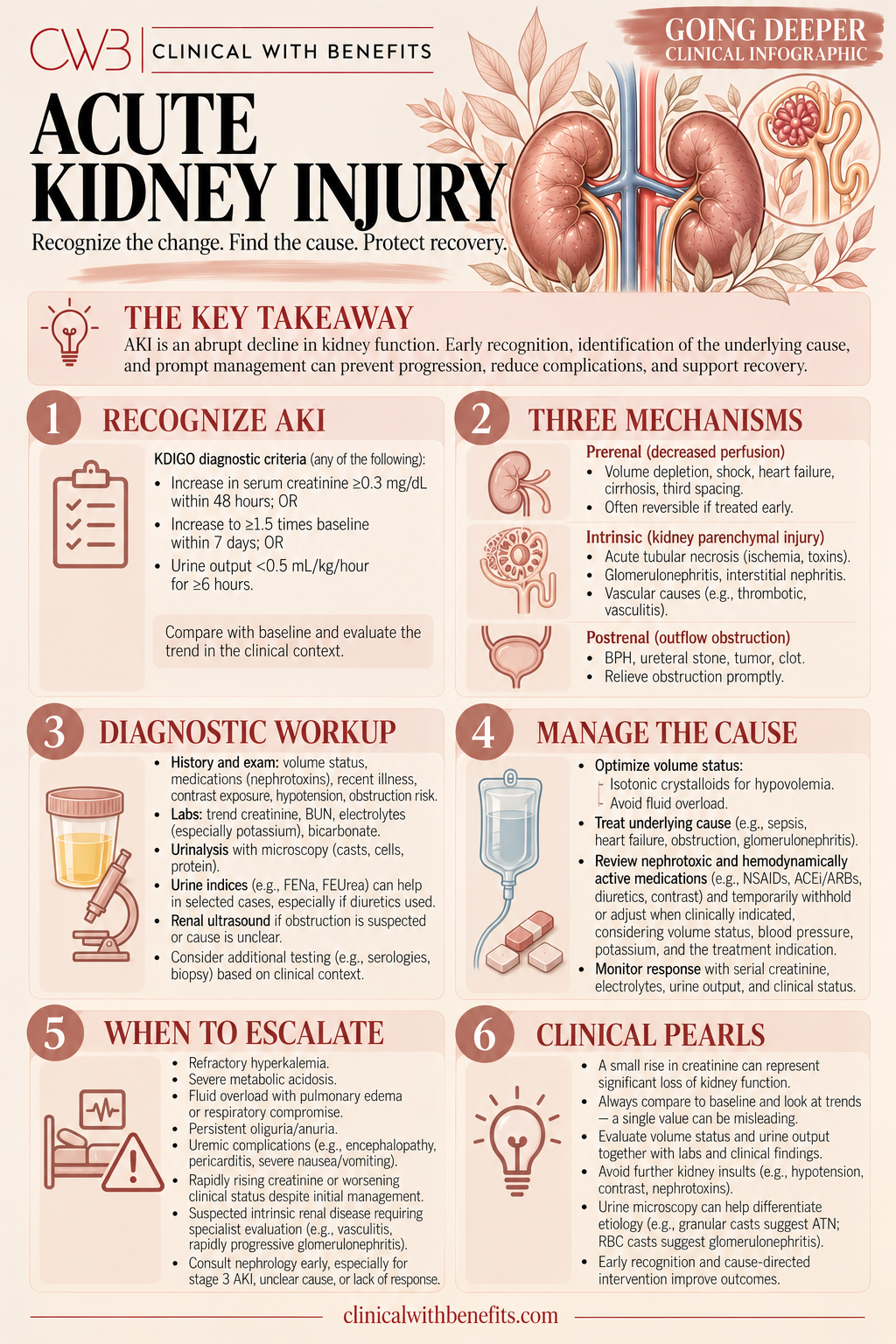
The three-category framework — pre-renal, intrinsic, post-renal — is the starting point. Most AKI in hospitalized patients is pre-renal or ischemic ATN, but obstruction is easy to miss and easy to reverse. Urine sediment and the medication list are two of the most informative and underutilized tools in the workup.
1 · Defining AKI — Trajectory, Not a Single Value
Acute kidney injury is defined by the KDIGO criteria: a rise in serum creatinine of 0.3 mg/dL or more within 48 hours, a rise to 1.5 times baseline or more within 7 days, or urine output below 0.5 mL/kg/hour for 6 or more hours. The key word is trajectory — AKI is not defined by a single creatinine value, but by a change from baseline over a defined time window.
This distinction matters clinically. A creatinine of 2.0 mg/dL in a patient whose baseline is 1.8 mg/dL is a very different situation from a creatinine of 2.0 mg/dL in a patient whose baseline is 0.8 mg/dL. Without a prior creatinine for comparison, the severity and acuity of the injury cannot be accurately assessed. Obtaining prior laboratory values is one of the first steps in any AKI workup.
Serum creatinine lags behind injury. It does not rise until roughly 50% of kidney function is lost, and it continues to rise for hours to days after the initial insult. A creatinine that is stable or only mildly elevated may still represent significant ongoing injury in the right clinical context. Urine output is often the earlier signal — oliguria frequently precedes the creatinine rise.
2 · The Three Categories — Pre-Renal, Intrinsic, Post-Renal
The organizing framework for AKI is the three-category model: pre-renal (underperfusion of intact kidneys), intrinsic (parenchymal kidney injury), and post-renal (obstruction to urine outflow). Most AKI in hospitalized patients is pre-renal or ischemic ATN — but the categories are not mutually exclusive, and mixed physiology is common.
Pre-renal AKI results from reduced effective renal perfusion. The kidneys are structurally intact but not receiving adequate blood flow. Causes include volume depletion (hemorrhage, GI losses, poor intake), low cardiac output (heart failure, cardiogenic shock), and vasodilation (sepsis, hepatorenal syndrome, medications). The kidneys respond by maximally conserving sodium and water — urine sodium is low, urine osmolality is high, and the BUN/creatinine ratio is often elevated. Pre-renal AKI is reversible when the perfusion problem is corrected, but prolonged pre-renal physiology leads to ischemic ATN.
Intrinsic AKI involves structural injury to the kidney itself. The most common cause in hospitalized patients is acute tubular necrosis (ATN), which results from ischemia (prolonged pre-renal state, hypotension, sepsis) or nephrotoxins (aminoglycosides, vancomycin, contrast agents, NSAIDs, myoglobin). Other intrinsic causes include acute interstitial nephritis (drug-induced, most commonly from NSAIDs, beta-lactams, PPIs, and sulfonamides), glomerulonephritis, and vascular processes. Urine sediment is the most direct window into intrinsic injury.
Post-renal AKI results from obstruction to urine outflow at any level — from the renal pelvis to the urethra. It is the most reversible category and the easiest to miss. Causes include benign prostatic hyperplasia (the most common cause in older men), bladder outlet obstruction, bilateral ureteral obstruction (pelvic malignancy, retroperitoneal fibrosis), and a blocked urinary catheter. Bladder scan or renal ultrasound should be performed early in any unexplained AKI, particularly in older men or patients with pelvic pathology.
3 · Urine Studies — The Most Direct Window Into the Process
Urinalysis and urine sediment are among the most informative and underutilized tools in the AKI workup. A bland urinalysis — no protein, no blood, no casts — supports pre-renal physiology. An active sediment shifts the differential toward intrinsic injury.
Urine sediment findings and their implications: muddy brown granular casts are the hallmark of ATN — they represent sloughed tubular epithelial cells and are highly specific for tubular injury. RBC casts indicate glomerulonephritis — blood in the urine with casts, not just hematuria. WBC casts indicate acute interstitial nephritis or pyelonephritis. Hyaline casts are nonspecific and can be seen in pre-renal states, dehydration, and with diuretic use.
Urine electrolytes and indices provide supporting data but have important limitations. The fractional excretion of sodium (FeNa) is calculated as (urine Na × plasma Cr) / (plasma Na × urine Cr) × 100. A FeNa below 1% suggests pre-renal physiology — the kidneys are avidly retaining sodium. However, diuretics, CKD, contrast nephropathy, early ATN, and myoglobinuria can all produce a low FeNa with intrinsic injury. In patients on diuretics, the fractional excretion of urea (FeUrea) is more reliable — a FeUrea below 35% supports pre-renal physiology.
The BUN/creatinine ratio above 20 supports pre-renal physiology — increased urea reabsorption in the setting of reduced tubular flow. However, GI bleeding, high protein intake, corticosteroids, and catabolic states also elevate BUN disproportionately. No single index replaces clinical synthesis.
4 · Nephrotoxins — Review the Medication List
Medication review is a required component of every AKI workup. Nephrotoxic medications are among the most common contributors to AKI in hospitalized patients, and they are frequently overlooked or continued after AKI is identified.
NSAIDs inhibit prostaglandin synthesis, reducing afferent arteriolar dilation and decreasing GFR — particularly in states of reduced effective renal perfusion (volume depletion, heart failure, cirrhosis). ACE inhibitors and ARBs reduce efferent arteriolar tone, which is the primary mechanism maintaining GFR when afferent flow is reduced. The combination of NSAID + ACE inhibitor (or ARB) + volume depletion is synergistically nephrotoxic and a classic cause of acute-on-chronic kidney injury.
Aminoglycosides (gentamicin, tobramycin, amikacin) cause direct tubular toxicity — the injury is dose-dependent and cumulative. Trough-based monitoring is standard, but extended-interval dosing (once-daily aminoglycosides) reduces nephrotoxicity compared to traditional dosing. Vancomycin nephrotoxicity is potentiated by concurrent aminoglycoside use and by trough-based dosing — AUC-guided vancomycin dosing reduces nephrotoxicity compared to trough-based monitoring.
Contrast-associated AKI peaks at 48–72 hours after exposure and typically resolves within 7–10 days. A creatinine that continues to rise beyond 72 hours after contrast suggests a different or additional cause. Pre-hydration with isotonic saline reduces contrast-associated AKI risk in high-risk patients. The risk of contrast-associated AKI has been substantially revised downward in recent literature — withholding necessary contrast imaging due to AKI concern often causes more harm than the contrast itself.
5 · Fluid Management — Responsiveness, Not Volume
Fluid resuscitation is appropriate for pre-renal AKI caused by volume depletion — but it is not appropriate for all AKI. Aggressive fluid administration in ATN, cardiorenal syndrome, or hepatorenal syndrome can worsen outcomes by increasing venous congestion, worsening renal perfusion pressure, and contributing to pulmonary edema.
The goal is fluid responsiveness — the ability of the kidneys to improve function with volume restoration — not fluid administration as a default intervention. Assess volume status carefully before giving fluids: clinical exam (JVP, skin turgor, mucous membranes, orthostatic vitals), urine output response, and dynamic measures of fluid responsiveness (passive leg raise, pulse pressure variation in mechanically ventilated patients) all inform the decision.
Isotonic crystalloids (normal saline, lactated Ringer's) are the preferred resuscitation fluids in AKI. Large volumes of normal saline can cause hyperchloremic metabolic acidosis, which may worsen outcomes in AKI patients. Balanced crystalloids (lactated Ringer's, Plasma-Lyte) are associated with lower rates of AKI and renal replacement therapy in critically ill patients compared to normal saline in recent randomized trials.
Diuretics do not treat AKI and do not accelerate recovery — they convert oliguric AKI to non-oliguric AKI, which may improve fluid management but does not change outcomes. Diuretics are appropriate for managing volume overload in AKI, not for treating the AKI itself.
6 · Indications for Renal Replacement Therapy
Renal replacement therapy (RRT) — hemodialysis, continuous renal replacement therapy (CRRT), or peritoneal dialysis — is indicated for life-threatening complications of AKI that cannot be managed conservatively. The classic indications are captured by the mnemonic AEIOU: Acidosis (severe metabolic acidosis, pH below 7.1–7.15, not responsive to bicarbonate), Electrolytes (severe hyperkalemia not responsive to medical management), Intoxication (dialyzable toxins — methanol, ethylene glycol, lithium, salicylates), Overload (refractory volume overload causing pulmonary edema), and Uremia (uremic encephalopathy, uremic pericarditis, uremic bleeding).
The timing of RRT initiation in AKI without these absolute indications remains an area of active investigation. Two large randomized trials (STARRT-AKI and IDEAL-ICU) found no benefit to early RRT initiation compared to a watchful waiting strategy in critically ill patients with severe AKI. Current practice favors a conservative approach — initiating RRT when clear indications develop rather than prophylactically.
CRRT is preferred over intermittent hemodialysis in hemodynamically unstable patients because it provides slower, continuous solute and fluid removal without the hemodynamic shifts associated with intermittent dialysis. In stable patients, intermittent hemodialysis is equally effective and more efficient.
7 · Monitoring, Recovery, and Follow-Up
Most AKI in hospitalized patients is reversible with treatment of the underlying cause. Recovery is defined by return of creatinine toward baseline — typically within 7–14 days for pre-renal AKI and ischemic ATN, though severe ATN may take weeks to months. Patients who do not recover kidney function within 90 days meet the definition of CKD.
During the recovery phase, monitor creatinine and urine output daily. Restart held medications cautiously — ACE inhibitors, ARBs, and NSAIDs should not be restarted until creatinine has returned to or near baseline. Nephrotoxic antibiotics should be dose-adjusted for the current GFR throughout the recovery period.
AKI is an independent risk factor for CKD, cardiovascular events, and mortality. Patients with AKI requiring hospitalization should have nephrology follow-up within 90 days of discharge. Repeat creatinine at 3 months confirms recovery or identifies progression to CKD. Patients with AKI who develop CKD should be referred to nephrology for ongoing management.
Apply It · Patient Scenario
A 74-year-old man with a history of hypertension, type 2 diabetes, and CKD stage 3 (baseline creatinine 1.6 mg/dL) is admitted with 3 days of nausea, decreased oral intake, and decreased urine output. He takes lisinopril, metformin, ibuprofen (for knee pain), and furosemide. Current creatinine is 3.1 mg/dL. Urine output over the past 8 hours is 80 mL. Urinalysis shows muddy brown granular casts. Blood pressure is 102/64 mmHg.
What is the most important immediate step?
A. Start IV fluids aggressively — the low blood pressure and decreased urine output indicate pre-renal AKI
B. Hold the ibuprofen, lisinopril, furosemide, and metformin; assess volume status carefully before giving fluids; the muddy brown casts indicate ATN
C. Order a renal ultrasound — the most likely cause is obstruction given his age
D. Start empiric antibiotics — the urinalysis findings suggest pyelonephritis
ANSWER
B. Hold the nephrotoxins and assess volume status carefully — the muddy brown casts indicate ATN, not pure pre-renal AKI.
RATIONALE
This patient has the classic NSAID + ACE inhibitor + volume depletion nephrotoxic triad. The ibuprofen reduces afferent arteriolar dilation; the lisinopril reduces efferent arteriolar tone; and decreased oral intake has created volume depletion. The furosemide is worsening the volume depletion. The muddy brown granular casts confirm that ATN has already developed — this is no longer purely pre-renal.
Aggressive IV fluids in a patient with ATN and CKD can cause volume overload without improving kidney function. The correct approach is to hold all nephrotoxins immediately (ibuprofen, lisinopril, furosemide), hold metformin (contraindicated in AKI due to lactic acidosis risk), carefully assess volume status, and give cautious IV fluid resuscitation if the patient is genuinely volume depleted. Monitor creatinine and urine output closely.
Obstruction is worth considering in an older man, but the clinical picture — nephrotoxic triad, muddy brown casts, and hemodynamic changes — points to ATN as the primary process. A bladder scan is reasonable but not the most important immediate step.
Clinical Pearl: The NSAID + ACE inhibitor + volume depletion triad is synergistically nephrotoxic. When you see this combination in a patient with AKI, hold all three before anything else.
NOW CHANGE ONE DETAIL
Same patient. Same presentation. Now the urinalysis shows no casts — just trace protein and a few hyaline casts. Blood pressure is 88/54 mmHg. He has been vomiting for 3 days with minimal oral intake.
UPDATED REASONING
Without muddy brown casts and with a bland urinalysis, the picture is more consistent with pre-renal AKI — the kidneys are underperfused but not yet structurally injured. The hypotension and vomiting support volume depletion as the primary driver. In this scenario, cautious IV fluid resuscitation with isotonic crystalloid is appropriate, with close monitoring of blood pressure, urine output, and creatinine response.
The nephrotoxins (ibuprofen, lisinopril, furosemide) should still be held — they are contributing to the AKI even in a pre-renal picture. The lesson: urine sediment is the key discriminator between pre-renal and intrinsic AKI when the clinical picture is ambiguous.
Understand It · The Nuance
The most common errors in AKI management are treating all AKI with aggressive fluids, missing obstruction, continuing nephrotoxins, and failing to recognize that creatinine lags behind injury.
AKI is a syndrome, not a diagnosis
Acute kidney injury is defined by trajectory — a rise of 0.3 mg/dL in 48 hours or 1.5x baseline in 7 days. The creatinine value alone does not tell you the cause, the severity of injury, or the prognosis. The clinical work is determining why the kidneys are injured.
The three categories frame the workup
Pre-renal (underperfusion), intrinsic (parenchymal injury), and post-renal (obstruction) are the organizing framework. Most AKI in hospitalized patients is pre-renal or ischemic ATN — but obstruction is easy to miss and easy to reverse if caught early.
Creatinine lags behind injury
Serum creatinine does not rise until roughly 50% of kidney function is lost. A 'normal' creatinine in a patient with low muscle mass, recent fluid resuscitation, or early injury does not rule out significant AKI. Urine output is often the earlier signal.
Urine studies are the most direct window into the process
Urinalysis and urine sediment can shift the differential quickly. Muddy brown granular casts indicate ATN. RBC casts indicate glomerulonephritis. WBC casts indicate interstitial nephritis. A bland urinalysis supports pre-renal physiology but does not rule out early intrinsic injury.
The medication list is a required part of the workup
NSAIDs, ACE inhibitors, ARBs, aminoglycosides, vancomycin, contrast agents, and diuretics are among the most common contributors to AKI in hospitalized patients. The NSAID + ACE inhibitor + volume depletion triad is synergistically nephrotoxic. Review and hold nephrotoxins early.
Fluid resuscitation is not always the answer
Aggressive fluid administration in a patient with ATN or cardiorenal syndrome can worsen outcomes. Fluid responsiveness — not fluid administration — is the goal in pre-renal AKI. Assess volume status carefully before giving fluids, and reassess after.
Clinical Pearl: Urine sediment is the most direct window into the AKI process. Muddy brown granular casts confirm ATN. A bland sediment supports pre-renal physiology. Do not skip the urinalysis in any AKI workup.
Clinical Pearls
Clinical Pearls
- Creatinine Lags Behind InjurySerum creatinine does not rise until roughly 50% of kidney function is lost. Urine output trends and clinical context often reveal AKI before the creatinine does.
- Always Rule Out ObstructionPost-renal AKI is the most reversible category and the easiest to miss. Bladder scan or renal ultrasound should be considered early, especially in older men, patients with pelvic malignancy, or unexplained AKI.
- Urine Sediment Is High YieldMuddy brown granular casts = ATN. RBC casts = glomerulonephritis. WBC casts = interstitial nephritis. A bland sediment supports pre-renal physiology but does not rule out early intrinsic injury.
- Hold Nephrotoxins EarlyNSAIDs, ACE inhibitors, ARBs, aminoglycosides, and contrast agents should be reviewed and held when AKI is identified. The NSAID + ACE inhibitor + volume depletion triad is synergistically nephrotoxic.
- FeNa Has LimitationsFeNa <1% supports pre-renal physiology in most contexts — but diuretics, CKD, contrast nephropathy, and early ATN can all produce a low FeNa with intrinsic injury. FeUrea is more reliable in diuretic-treated patients.
- Fluid Responsiveness Is the GoalAggressive fluid administration in ATN or cardiorenal syndrome can worsen outcomes. Assess volume status carefully before giving fluids and reassess after. The goal is fluid responsiveness, not fluid administration.
Bottom Line
AKI workup begins with the three-category framework — pre-renal, intrinsic, post-renal — and requires a prior creatinine for context. Urine sediment and the medication list are the two most informative tools. Hold nephrotoxins early, assess volume status before giving fluids, rule out obstruction, and monitor for the life-threatening complications that require renal replacement therapy.
EVIDENCE & REFERENCES
- KDIGO Clinical Practice Guideline for Acute Kidney Injury. Kidney Int Suppl. 2012;2(1):1-138. doi:10.1038/kisup.2012.1
- Kellum JA, et al. Diagnosis, evaluation, and management of acute kidney injury: a KDIGO summary. Crit Care. 2013;17(1):204. doi:10.1186/cc11454
- Perazella MA, Coca SG. Traditional urinary biomarkers in the assessment of hospital-acquired AKI. Clin J Am Soc Nephrol. 2012;7(1):167-174. doi:10.2215/CJN.09490911
- Mehta RL, et al. Spectrum of acute renal failure in the intensive care unit: the PICARD experience. Kidney Int. 2004;66(4):1613-1621. doi:10.1111/j.1523-1755.2004.00927.x